Toluene is the major component of organic industrial solvents and the primary neurotoxic agent in paint thinners, lacquers, and spray paints. As a lipid-soluble aromatic hydrocarbon, it crosses the blood-brain barrier with ease and accumulates in lipid-rich brain structures. Modern neuroimaging has revealed the devastating structural consequences of chronic exposure — consequences that were once invisible but are now unmistakably documented.
paint-and-liquid-coatings-risks
Toluene Brain Damage: What MRI Reveals About Chronic Paint Solvent Exposure
Sundial Research Team·January 25, 2025·6 min
MRI studies of chronic toluene abusers — who represent an extreme exposure scenario — reveal a characteristic triad of abnormalities:
Ready to Start Your Project?
From one-off customs to 15,000-part production runs — get precise pricing in 24 hours.
On This Page
Toluene Brain Damage: What MRI Reveals About Chronic Paint Solvent Exposure
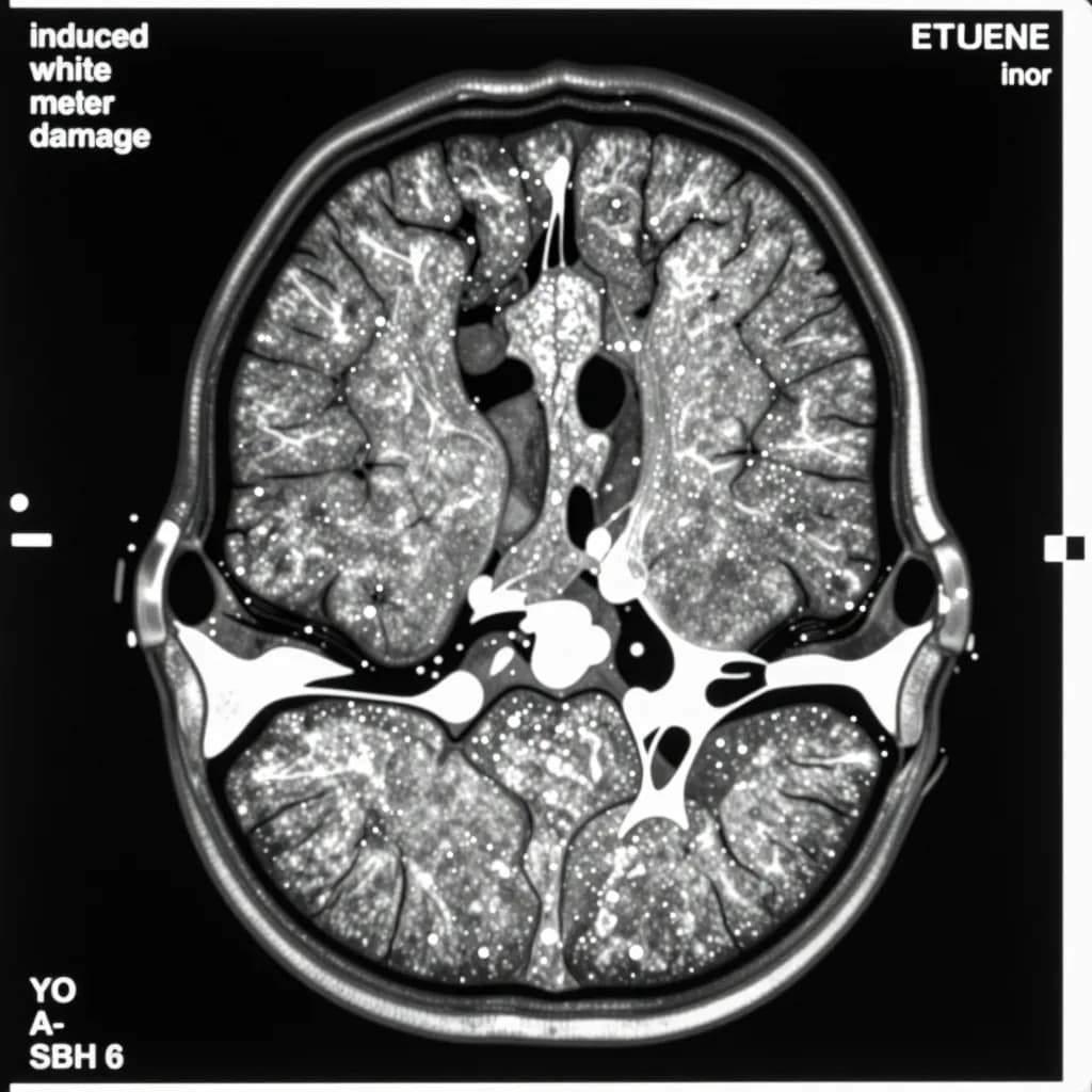
The Structural Brain Damage Pattern
| Abnormality | Prevalence | Significance |
|---|---|---|
| White matter lesions | 46% | Demyelination and gliosis |
| Atrophic ventricular/sulcal dilatation | 27% | Generalized cerebral atrophy |
| Thalamic hypointensity | 20% | Iron deposition from axonal loss |
Both duration >4 years was significantly associated with white matter changes (P<0.05) and thalamic hypointensity (P<0.01).
White Matter: The Primary Target
White matter changes are the hallmark of chronic toluene neurotoxicity. The pathology starts in the deep periventricular white matter and spreads into peripheral white matter with continued exposure. Histologically, these changes represent:
- Demyelination: Loss of the lipid-rich myelin sheath that insulates axons
- Gliosis: Scarring and proliferation of astrocytes in response to injury
- Axonal degeneration: Breakdown of the nerve fibers themselves
The correlation between white matter damage and cognitive impairment is strong. In a study of 14 chronic toluene abusers, the degree of white matter abnormality was strongly correlated (p<0.01) with neuropsychological impairment. Eight of 14 were classified as cognitively impaired.
Thalamic Hypointensity and Iron Deposition
Thalamic hypointensity on MRI — appearing as dark spots in the thalamus — is a distinctive finding in chronic toluene exposure. The mechanism is iron deposition resulting from demyelination and axonal loss. When myelin breaks down, iron — normally bound in myelin — is released and deposits in surrounding tissue, creating the characteristic signal changes.
This finding is important because the thalamus serves as a relay station for sensory and motor signals, as well as a regulator of consciousness and sleep. Thalamic damage contributes to the cognitive and psychiatric symptoms of chronic solvent encephalopathy.
Occupational Painter Studies
While toluene abusers represent extreme exposure, occupational painters show similar patterns at lower severity:
Arlien-Soborg (1979): The Landmark Study
In 70 house painters referred for suspected organic solvent intoxication, neuroradiological examination (PEG or CT) demonstrated cerebral atrophy in 31 of 50 cases with no competing etiology. Neuropsychological examination showed intellectual impairment in 39 of 50.
Keski-Santti (2009): MRI in CSE Patients
MRI scans of 71 chronic solvent encephalopathy patients found abnormalities in 38%:
- Brain atrophy in any area: 24%
- Abnormal white matter hyperintensities: 28%
- Cerebral and cerebellar atrophy correlated with duration of exposure
Feldman (1999): Case Study
MRI of a 57-year-old painter with 30+ years of solvent exposure revealed global and symmetrical volume loss, involving more white than gray matter. Serial testing after exposure cessation showed persistent cognitive deficits without progression — the classic non-progressive course of CSE.
fMRI: Functional Consequences
Beyond structural damage, functional neuroimaging reveals how solvent exposure alters brain activity. An fMRI study comparing 27 solvent-exposed industrial painters with 27 controls during N-back working memory tasks found:
- Significantly worse performance on working memory tests
- Lower task-evoked activation in anterior cingulate cortex
- Reduced activation in dorsolateral prefrontal cortex
- Decreased parietal activation — regions serving attention and working memory
Lifetime solvent exposure was negatively correlated with activation in these regions: more exposure, less brain activity.
Cerebellar Damage
Toluene-induced chronic toxic encephalopathy causes characteristic cerebellar symptoms including:
- Ataxia (unsteady gait)
- Tremors
- Nystagmus (involuntary eye movements)
- Cognitive impairment and psychiatric disorders
- Temporal lobe epilepsy (incidence of 13.6% in one study)
The cerebellum — historically viewed as solely responsible for motor coordination — is now recognized as critically involved in cognitive and emotional processing. Cerebellar damage contributes to the broad symptom profile of CSE.
Mechanism: Why the White Matter?
Organic solvents preferentially damage white matter for several reasons:
- Lipid solubility: White matter is lipid-rich; solvents partition into myelin
- High surface-area-to-volume ratio: Myelinated axons present extensive membrane targets
- Metabolic vulnerability: Oligodendrocytes (myelin-producing cells) are metabolically active and susceptible to oxidative stress
- Axonal transport disruption: Solvents interfere with critical transport mechanisms
Reversibility: The Critical Window
The structural damage documented on MRI is largely irreversible. While mild Type 1 symptoms may resolve with early exposure cessation, established white matter damage and cerebral atrophy persist. The Bruhn (1981) 2-year follow-up of 26 house painters found that neurological status, neuropsychological impairment, and cerebral atrophy did not change significantly after exposure cessation.
The message for prevention is clear: intervention must occur before structural damage develops. Once MRI-visible changes appear, the window for meaningful recovery has closed.
Powder Coating: Eliminating the Exposure Pathway
Powder coatings contain no toluene and no organic solvents. The elimination of solvent carriers removes the primary neurotoxic exposure pathway. For facilities where worker neurological health is a priority, powder coating is not merely a safer alternative — it is the only alternative that can prevent the structural brain damage documented in decades of neuroimaging research.
Ready to Start Your Project?
From one-off customs to 15,000-part production runs — get precise pricing in 24 hours.